Boston Terrier Cured Thanks to Groundbreaking 3D Laparoscopic Procedure
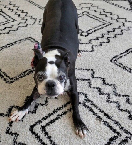
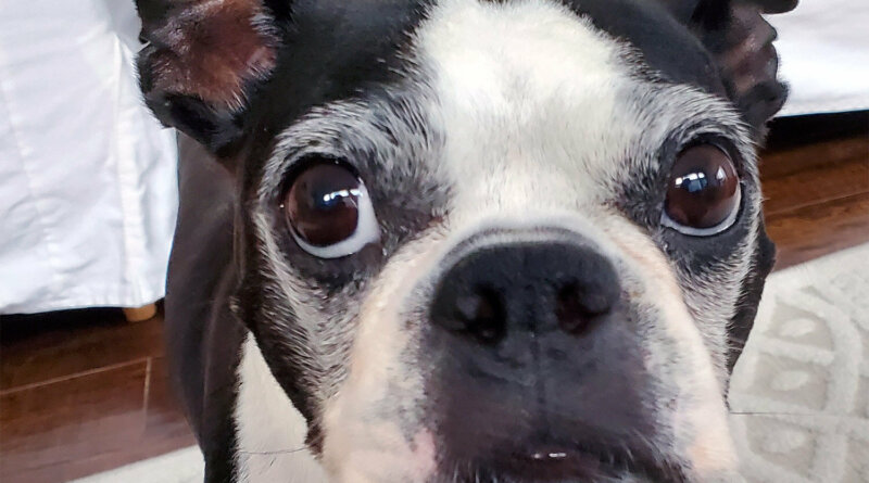
Louie, an 8-year-old male Boston terrier, was diagnosed with Cushing’s disease by his primary veterinarian. Cushing’s disease causes a dog’s adrenal glands to produce too much cortisol, a chemical that controls many aspects of a dog’s body, including its weight, its ability to fight infections, maintain blood sugar levels, and many other vital functions. In Louie, the Cushing’s disease was caused by a tumor in his right adrenal gland. His primary veterinarians referred Louie to the Soft Tissue Surgery Service at the UC Davis veterinary hospital for surgical removal of his right adrenal gland.
As a pioneer in minimally invasive procedures, UC Davis has been performing laparoscopic surgeries for many years, including adrenalectomies. Normally, these surgeries produce an on-screen two-dimensional (2D) image from a camera in the scope. Surgeons are able to navigate the animal’s body proficiently after a learning curve, however they lose depth perception with a 2D image which may lead to surgical errors or prolong surgical time.
Louie recovered well from a groundbreaking surgery to remove an adrenal gland at the UC Davis veterinary hospital.
Recently, UC Davis acquired a three-dimensional (3D) scope that greatly improves on the 2D shortcomings. With two cameras in the scope instead of one, two images are presented on-screen. When viewed with 3D glasses, the two images are fused together due to the polarization of the glasses, creating a 3D image on the monitor, helping veterinarians to better navigate their surgical field.
GET THE BARK NEWSLETTER IN YOUR INBOX!
Sign up and get the answers to your questions.
While this technology has been used in human surgeries for several years, it is new technology for veterinary medicine. UC Davis veterinarians believe Louie’s procedure to be the first laparoscopic adrenalectomy performed on an animal utilizing 3D technology. Studies on human procedures have shown 3D surgeries to decrease surgical time and decrease surgical errors, and the school’s faculty hope to see those advantages realized for their veterinary patients also.
“I hope this allows us to push the envelope for different types of surgeries that we could consider performing minimally invasively,” said Louie’s surgeon, Dr. Ingrid Balsa. “I think this will also provide a stepping stone for resident training in regards to learning laparoscopic procedures, which have different instrumentation and techniques compared to traditional open surgeries. The 3D scope will remove one barrier, loss of depth perception, in learning laparoscopy.”

Louie’s procedure was made possible due to the generous financial support of Mary Kariotis and the UC Davis Center for Companion Animal Health Equipment Fund that allowed the hospital to purchase the 3D laparoscopic equipment. Kariotis, a Bay Area businesswoman and philanthropist, became interested in helping UC Davis after Dr. Philipp Mayhew and the Soft Tissue Surgery Service saved her dog with a similar surgery to remove one of his adrenal glands after a tumor was discovered.
“Dr. Mayhew changed his vacation plans to accommodate my dog’s critical surgery,” said Kariotis. “It seems every veterinarian I come in contact with has been taught by and/or holds great admiration for Dr. Mayhew.”
Dr. Balsa agrees, having completed her American College of Veterinary Surgeons fellowship in minimally invasive surgery in 2020 under Mayhew’s mentorship. Dr. Mayhew also played a large part in mentoring Dr. Balsa through her three-year surgical residency, completed at UC Davis in 2016.
“Pets are an integral part of our families that we love as our children,” said Kariotis, expressing her deep affection for animals. “I will be forever grateful for the care we received at UC Davis, which is unsurpassed by any other veterinary clinic in a myriad of ways. I hope my gift can make similar surgeries as successful as possible and help all our pets live long lives.”
The tumor on Louie’s right adrenal gland was benign, and his left adrenal gland is now performing normally after an initial steroid supplementation. So, his veterinarians consider his Cushing’s disease to be cured. His owners Aaron and Ivonne Jamison reports that Louie’s energy level is excellent, and his appetite and thirst are back to normal.
The Jamisons concur with Kariotis’ assessment of animals’ roles in our lives, as well as the care provided at UC Davis.
“Working with Dr. Balsa and her team was excellent right from the start,” said Aaron. “Everyone was engaged, enthusiastic, and compassionate in the way they treated Louie, as well as us. Understandably, Ivonne and I were anxious about our baby undergoing major surgery. We have no children, so Louie is the focus of our parental instincts. We had a lot of reservations about an adrenalectomy, especially since most cases in the past have been handled open as opposed to laparoscopically. However, thanks to some research, we discovered that UC Davis has pioneered the laparoscopic approach, with great success. If Louie ever needs a major procedure in the future, we will be returning to UC Davis – no question about it.”
Louie’s surgery showcases an example of groundbreaking surgical procedures that will be a hallmark of the future Veterinary Medical Center at UC Davis. As the final phase of the VMC, an entirely new Small Animal Hospital will be constructed, increasing the size and scope of the current facility. This will allow clinicians to expand their innovative procedures and continue to push the limits of veterinary medicine.


