Help for Mast Cell Tumor Patients – Dogster
It’s a diagnosis no veterinarian likes to make. As soon as the word is spoken, it shatters the room. This time was no different. “It’s cancer. Max has a malignant mast cell tumor. I am so sorry.”
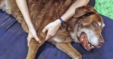
The mother of two seated across from me immediately began sobbing. The handsome Labrador Retriever lying at her feet looked up worriedly. As I offered her tissues, I reflected on how we got here and where we were headed. None of us knew a miracle was waiting.
What Is a Mast Cell Tumor?
Mast cell tumors (MCT) are the most common skin tumors in dogs. Research shows MCTs account for about 20% of all skin cancers. MCTs are so common, I teach my young vets to presume any lump or bump on or just underneath the skin is a mast cell tumor until proven otherwise. Scientists don’t understand why this cancer occurs in dogs, but mast cells are made in the bone marrow and travel to sites in response to inflammation. Fortunately, most MCTs are solitary and don’t metastasize or spread to other parts of the body. Only about 11 to 14% of dogs with an MCT will have more than one tumor. MCTs can appear anywhere on the body and usually are slow-growing, causing many dog owners to overlook or forget about the mass until it’s too late. As with many cancers, when diagnosed early and small, MCTs have a better prognosis.
Mast cell tumors are more common in older dogs, typically between ages 7 and 9. Labrador and Golden Retrievers, Shar-Peis, Boxers, Boston Terriers, American Staffordshire Terriers, Pugs and French Bulldogs are some breeds more likely to develop MCTs, although any dog at any age can develop them.
How to Know if It Is an MCT
Max fit squarely into the MCT risk matrix, so when his mom brought him that day after the sudden appearance of a peasized growth near the top of his rear paw, we went into action. Any time a dog has a movable skin tumor, the first thing I do is a fine-needle aspiration (FNA). Because MCTs produce large numbers of mast cells in a confined space, it’s relatively easy to determine if a mass is an MCT or not. In cases of previous MCT, or if I’m particularly concerned, pretreatment with diphenhydramine (Benadryl) can help reduce the risk of swelling and inflammation after the FNA.
The procedure is straightforward: A needle is carefully inserted into the tumor and cells suctioned out. The sample is placed on a slide for staining and histopathological evaluation. I prefer to do an initial evaluation of the slide in my clinic. If I see mast cells or other suspicious tissues, I refer the test to a veterinary pathologist. When I peered into Max’s slide, I was met by a wall of characteristic small- to medium-sized round, purple-red cells consistent with mast cells. We overnighted the test.
The next day the lab confirmed the diagnosis. We performed regional lymph node FNAs along with chest X-rays to check for potential spread and blood tests. If there was a time for a miracle, it was now.
Time for That Miracle
Max’s tests showed no signs of spread. Historically, MCTs would be surgically removed with a broad border to prevent recurrence. The challenge with many dogs, including Max, was that the rear leg didn’t offer much depth beneath the cancer or extra skin to close a large excision. In these cases, radiation therapy may be required after surgery. But that was 2020.
It was 2021, and a new MCT treatment had just been approved in the United States for canine non-metastatic MCT. I had been reading reports from other countries about Stelfonta and was eager to see it in action. Because it was so new, I referred Max to an oncologist for treatment. The great news is any veterinarian can administer Stelfonta, and it’s becoming widely available. It’s also affordable, especially when compared to surgery and follow-up care.
Let’s Talk Stelfonta
Tigilanol tiglate injection, sold under the brand name Stelfonta, was discovered in the Australian rainforest blushwood plant (Fontainea picrosperma). It was approved by the FDA for treating non-metastatic mast cell tumors in dogs in November 2020 and is only available through a veterinarian.
It’s injected directly into the tumor and literally kills only the tumor cells, leaving surrounding tissues unharmed. The tumor slowly dissolves, forming what looks like an open sore, over the next few weeks. Studies prove about 75% of MCTs are removed with a single injection, and 88% with two doses. Sounds like a miracle to me.
Recovery Time
And it was. Max received his injection and within a week the cancer was turning into what can only be described as “mush.” The drug maker instructs dog owners to allow the dog to lick and clean it (no E-collars!), and not bandage or cover the wound. Incredibly, the drug also promotes healing of normal tissues, so no antibiotics are needed.
Within two months, Max’s tumor site was completely healed with minimal scarring. Because the treatment is relatively new and we don’t understand what causes MCTs in the first place, it’s too early to say if dogs like Max will suffer from future tumors.
This treatment is best for small, superficial cancers that haven’t spread. Not all MCTs can be treated with Stelfonta, and your veterinarian will determine if your dog is an appropriate candidate. The sloughing, open wound can be unsettling for some, so be prepared to observe a large sore for a few weeks. It took me a minute to subdue my veterinary-instinct to bandage and prescribe.
Max got his miracle. During our last follow-up visit a couple of months post-Stelfonta, I realized the miracle was also for us. Max’s mom was jubilant and remarked she was taking advantage of every day she had with Max and her human family. Overcoming the “word we hate to hear” had given her renewed appreciation for the simple joys of life and time spent with loved ones. Now that’s a true miracle.


